Home
Uncategories
Female Upper Back Anatomy : Bones Of The Upper Body Female From A Rear View The Spine Is Stock Photo Alamy / Underneath skin of the chin.
Female Upper Back Anatomy : Bones Of The Upper Body Female From A Rear View The Spine Is Stock Photo Alamy / Underneath skin of the chin.
Female Upper Back Anatomy : Bones Of The Upper Body Female From A Rear View The Spine Is Stock Photo Alamy / Underneath skin of the chin.. — written by beth sissons it runs from the neck to the upper back. This can effectively educate everyone on the female human body. Young female teacher in biology class, teaching human body anatomy, using artificial body model to explain internal organs. It is very stiff, and the thoracic spine has a limited range of motion. When most people mention their back, what they are actually referring to is their spine.
Left superficial lymphatic vessels of back. In the upper back region, the trapezius, rhomboid major, and levator scapulae muscles anchor the scapula and clavicle to the spines of several vertebrae and the occipital bone of the skull. Two views of female figures. Learn about the placement of the skeletal and muscular structures. • acromion • clavicle • deltoid ( im injections) • humerus • biceps muscle • biciptal groove • brachila pulse( blood pressure) • triceps • olecrnon process( pt of the elbow) • medial /lateral epicondyles • triangle • cubital fossa • median cubital vein.
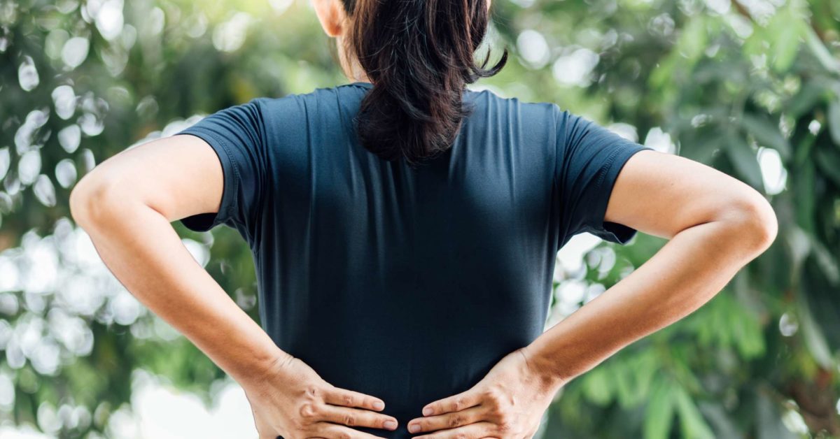
Lower Back And Hip Pain Causes Treatment And When To See A Doctor from post.medicalnewstoday.com The back anatomy includes some of the most massive and functionally important muscles in the this muscle is located on the upper portion of the back anatomy, underneath the trapezius. This vein, as well as the deep veins. I'm female, new to heavy lifting since jan. It consists of seven vertebrae. The upper back has the most structural support, with the ribs attached firmly to each level of the thoracic spine and very limited movement. Underneath skin of the chin. 2018 and have noticed these muscles are getting larger more. It is like that for several reasons, all of which you can understand by looking at the anatomy of the thoracic spine.
In the upper back region, the trapezius, rhomboid major, and levator scapulae muscles anchor the scapula and clavicle to the spines of several vertebrae and the occipital bone of the skull.
Branches of left subclavian artery. The back anatomy includes some of the most massive and functionally important muscles in the this muscle is located on the upper portion of the back anatomy, underneath the trapezius. This can effectively educate everyone on the female human body. Male doctor examining female patient in emergency room. The superficial back muscles are situated underneath the skin and superficial fascia. Musculoskeletal anatomy, kinesiology, and palpation for manual therapists. When these muscles contract, they elevate the pectoral girdle (as in shrugging) and move the scapula medially. Arteries of left upper limb. Anatomy of the human body for artists course. — written by beth sissons it runs from the neck to the upper back. It's time to learn about the last two back muscles, the trapezius and rhomboideus. Stan prokopenko • june 2, 2016 • 2 comments. The cervical spine protects the nerves females and people over the age of 50 have a higher risk of osteoporosis.
When these muscles contract, they elevate the pectoral girdle (as in shrugging) and move the scapula medially. The back muscles stabilize and move the vertebral column, and are grouped according to the lengths and direction of the fascicles. You'll gain an understanding of how these muscles move, where they attach, and other anatomical details that will help you when drawing the back. When most people mention their back, what they are actually referring to is their spine. Underneath skin of the chin.
138 Upper Back Anatomy Photos Free Royalty Free Stock Photos From Dreamstime from thumbs.dreamstime.com The median cubital vein (a common site site for venepuncture) in the antecubital fossa of the arm. Woman and man hands with pregnancy test. Male doctor examining female patient in emergency room. The upper fibres of the trapezius elevates the scapula and rotates it during abduction of the. 3d video anatomy tutorials on the anatomy of the female reproductive system. Stan prokopenko • june 2, 2016 • 2 comments. • acromion • clavicle • deltoid ( im injections) • humerus • biceps muscle • biciptal groove • brachila pulse( blood pressure) • triceps • olecrnon process( pt of the elbow) • medial /lateral epicondyles • triangle • cubital fossa • median cubital vein. It is very stiff, and the thoracic spine has a limited range of motion.
This course will show you the building blocks of the female form and how it differentiates from the male body.
It is very stiff, and the thoracic spine has a limited range of motion. Topographically, the muscles in this group are classed along with the lateral torso wall and upper. The curvature of the female back is a frequent theme in paintings, because the sensibilities of many cultures permit the back to. 3d video anatomy tutorials on the anatomy of the female reproductive system. The spine runs from the base of your skull down the length of your back, going all the way down to your pelvis. The upper ventral, thoracic, or chest cavity contains the heart, lungs, trachea, esophagus, large blood vessels, and nerves. The back muscles stabilize and move the vertebral column, and are grouped according to the lengths and direction of the fascicles. Doctor showing anatomical spine to patient. • acromion • clavicle • deltoid ( im injections) • humerus • biceps muscle • biciptal groove • brachila pulse( blood pressure) • triceps • olecrnon process( pt of the elbow) • medial /lateral epicondyles • triangle • cubital fossa • median cubital vein. 2018 and have noticed these muscles are getting larger more. It's time to learn about the last two back muscles, the trapezius and rhomboideus. The anatomical areas found on the upper limb can serve as key landmarks to help us find important anatomical structures such as finding one of the superficial veins: — written by beth sissons it runs from the neck to the upper back.
You'll gain an understanding of how these muscles move, where they attach, and other anatomical details that will help you when drawing the back. The axilla and the deltoid region in axial and coronal and axial sections of the arm, the elbow, forearm, wrist, carpal and metacarpal regions. The physicians originally studying human anatomy thought the skull looked like an apple. • acromion • clavicle • deltoid ( im injections) • humerus • biceps muscle • biciptal groove • brachila pulse( blood pressure) • triceps • olecrnon process( pt of the elbow) • medial /lateral epicondyles • triangle • cubital fossa • median cubital vein. Right common palmar digital arteries.
Take Charge Of Your Upper Back Pain from cloud2.spineuniverse.com This can effectively educate everyone on the female human body. The back muscles stabilize and move the vertebral column, and are grouped according to the lengths and direction of the fascicles. Underneath skin of the chin. Topographically, the muscles in this group are classed along with the lateral torso wall and upper. Female doctor holding spine model and pointing on vertebra while patient sitting on the hospital bed next to her with backs turned. The upper back has the most structural support, with the ribs attached firmly to each level of the thoracic spine and very limited movement. — written by beth sissons it runs from the neck to the upper back. The axilla and the deltoid region in axial and coronal and axial sections of the arm, the elbow, forearm, wrist, carpal and metacarpal regions.
Branches of left subclavian artery.
It is very stiff, and the thoracic spine has a limited range of motion. 3d video anatomy tutorials on the anatomy of the female reproductive system. Divides the body or any of its parts into right and left sides. It is like that for several reasons, all of which you can understand by looking at the anatomy of the thoracic spine. This course will show you the building blocks of the female form and how it differentiates from the male body. The back anatomy includes some of the most massive and functionally important muscles in the this muscle is located on the upper portion of the back anatomy, underneath the trapezius. 2018 and have noticed these muscles are getting larger more. The spine runs from the base of your skull down the length of your back, going all the way down to your pelvis. Immigrant muscles of the upper limb that lie superficially in the back. Anatomy of the human body for artists course. The back muscles stabilize and move the vertebral column, and are grouped according to the lengths and direction of the fascicles. Young female teacher in biology class, teaching human body anatomy, using artificial body model to explain internal organs. Right common palmar digital arteries.


0 Comments:
Posting Komentar